
Glutathione vs NAD+: Two Redox Molecules, Different Jobs
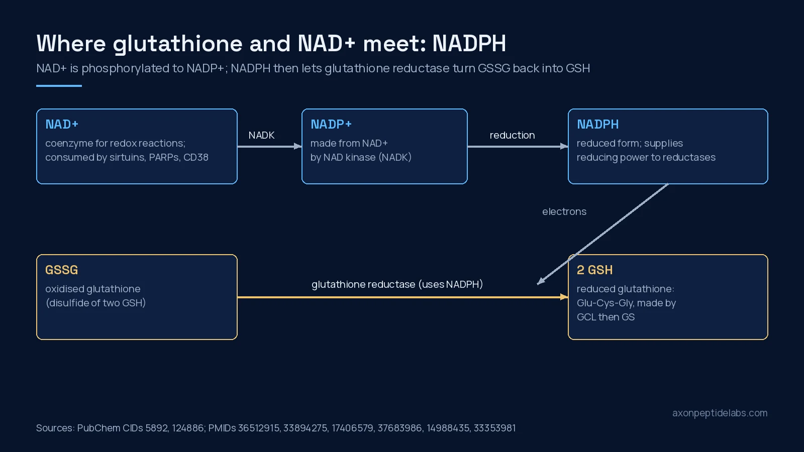
Short answer: glutathione and NAD+ are both central to cellular redox chemistry, but they are different kinds of molecule doing different jobs. Glutathione (GSH) is a tripeptide, γ-glutamyl-cysteinyl-glycine, and the most abundant low-molecular-weight thiol in animal cells; with its oxidised form (GSSG) it is the cell’s main redox buffer [1]. NAD+ is a dinucleotide coenzyme that carries electrons in metabolic reactions and is also used up as a substrate by sirtuins, PARPs and CD38 [2][3]. They connect through NADPH: NAD+ is phosphorylated to NADP+ [4], and NADPH is what glutathione reductase uses to turn GSSG back into GSH [5][6].
For laboratory research only. Not for human or animal use. This article gives no dosing, administration or therapeutic advice.
For anyone comparing the two as research materials, the more useful questions are what each molecule is, what happens to it outside a cell, and what controlled human studies have and have not found. Those are the sections below, with the null results kept in.
Identity
Identifiers were read from PubChem on October 1, 2026. Note that NAD+ is not a peptide, even though it is commonly sold alongside peptides.
| Item | Glutathione (reduced, GSH) | NAD+ (nadide) |
|---|---|---|
| Class | Tripeptide thiol (γ-Glu-Cys-Gly; the γ-linkage is unusual) | Dinucleotide coenzyme (nicotinamide and adenine nucleotides joined by a pyrophosphate) |
| Molecular formula | C10H17N3O6S | C21H27N7O14P2 |
| Molecular weight | 307.33 | 663.4 |
| PubChem CID | 124886 | 5892 |
| CAS (PubChem synonyms) | 70-18-8 | 53-84-9 |
| Made in cells by | Glutamate-cysteine ligase (rate-limiting), then glutathione synthetase [7][6] | Several routes from tryptophan and vitamin B3 forms, including NR and NMN [2][8] |
| Redox partner | GSSG (oxidised disulfide) | NADH (reduced form); also phosphorylated to NADP+/NADPH [4] |
| Consumed by | Glutathione peroxidases and transferases; degraded outside cells by γ-glutamyl transferase [6] | Sirtuins, PARPs and CD38, which break it down [2][9] |
What each does inside a cell
Glutathione: the thiol buffer
The GSH/GSSG pair is described as the major redox couple in animal cells [1]. Much of glutathione’s antioxidant work is done through enzymes: glutathione peroxidases use it to remove hydrogen peroxide and lipid hydroperoxides, and glutathione transferases use it in detoxification [6]. Synthesis takes two ATP-dependent steps, with glutamate-cysteine ligase and cysteine supply both limiting [7][6]. Several reviews link lower cellular glutathione with ageing, attributed mainly to reduced glutamate-cysteine ligase activity, although the mechanisms are not settled [10][6].
NAD+: coenzyme and consumable substrate
NAD+ accepts electrons in glycolysis and the citric acid cycle and passes them on as NADH. Separately, sirtuins, PARPs and CD38 cleave NAD+ as part of their reactions, so cells have to keep remaking it [2][3]. In mice, CD38 expression and activity rose with age and CD38 was required for the age-related fall in tissue NAD+ [9]. In human pelvic skin from 49 people, NAD+ levels fell with age while PARP activity rose, more clearly in males [11].
Why NAD+ research moved to precursors
Much of the animal work that drew attention to NAD+ used smaller precursor molecules rather than NAD+ itself. In mice fed a high-fat diet, NAD+ synthesis through the NAMPT pathway was impaired in metabolic organs, and nicotinamide mononucleotide (NMN) restored tissue NAD+ and improved glucose tolerance [12]. A 2018 review of NAD-boosting molecules in animals frames the field the same way: restoring NAD+ in old or diseased animals, mostly via precursors, is the intervention studied [8]. That history explains why the human trials below mostly test NR or NMN, and why their results should not be read across to NAD+ supplied directly.
The meeting point
NAD kinase phosphorylates NAD+ to make NADP+ [4], which enzymes such as glucose-6-phosphate dehydrogenase and malic enzyme reduce to NADPH [13]. Glutathione reductase then uses NADPH to regenerate GSH from GSSG [5][6]. So the two pools are linked, but indirectly and through an enzyme chain: adding one is not the same as adding the other.
What happens to each outside a cell
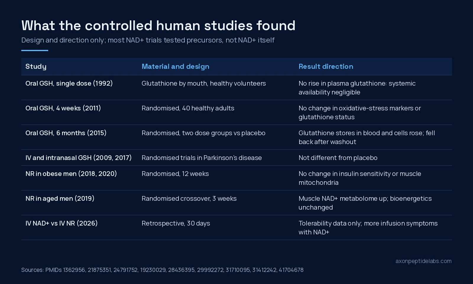
This is the practical difference most supplier pages skip. Glutathione is broken down outside cells by membrane-bound γ-glutamyl transferase [6]. In seven healthy volunteers, a single oral dose of glutathione did not raise plasma glutathione, cysteine or glutamate over the following four and a half hours; the authors judged systemic availability negligible, because of hydrolysis in gut and liver [14].
NAD+ is a large charged molecule. In cell experiments, prolonged exposure to extracellular NAD+ raised intracellular NAD+ at first, then cells “reset” to baseline by reducing uptake of NAD+ breakdown products, cutting expression of CD73, the NMN transporter SLC12A8 and NR kinase [15]. In mice, injected NAD+ did reach the hypothalamus, through a connexin 43-dependent route [16]. These findings come from cell lines and mice, and they point the same way: what reaches the inside of a cell depends on breakdown and transport, not just on the amount supplied.
Controlled human studies, by design
For glutathione, the human record is conflicting. A randomised four-week trial in 40 healthy adults found no change in urinary oxidative-stress markers or in glutathione status [17]. A six-month randomised trial in 54 non-smoking adults found that glutathione stores in blood and cells rose in both dose groups and returned to baseline after washout [18]. In Parkinson’s disease, a randomised pilot of intravenous glutathione found no significant difference from placebo on the rating scale [19], and a phase IIb trial of intranasal glutathione concluded it was not superior to placebo over three months, in part because the placebo group improved unusually well [20].
For NAD+, most controlled human trials did not use NAD+ at all; they used precursors such as nicotinamide riboside (NR). In 12 aged men, NR raised the muscle NAD+ metabolome without changing mitochondrial bioenergetics [21]. In a 12-week randomised trial in obese, insulin-resistant men, NR did not improve insulin sensitivity, energy expenditure or body composition [22], and muscle biopsies from the same programme showed no change in mitochondrial respiration or content [23]. A 2026 systematic review of 113 human and rodent studies found no eligible outcome trials of intravenous or intramuscular NAD+ itself and called clinical effectiveness inconclusive [24]. A retrospective 2026 comparison of intravenous NAD+ and NR reported tolerability only, with more infusion-related symptoms in the NAD+ group [25].
Side-by-side comparison
| Question | Glutathione | NAD+ |
|---|---|---|
| Main role | Thiol redox buffer; cofactor for peroxidases and transferases [1][6] | Electron carrier; substrate for sirtuins, PARPs, CD38 [2] |
| Fate outside the cell | Cleaved by γ-glutamyl transferase; oral single dose not systemically available [6][14] | Cleaved and taken up as smaller products; cells reset intracellular levels with prolonged exposure [15] |
| Age-related change | Lower cellular levels linked to reduced synthesis (reviews) [10] | Lower in some tissues; evidence limited and tissue-specific [11][26] |
| Best human data | Oral trials with conflicting results [17][18] | Precursor (NR) trials; none of NAD+ itself with clinical outcomes [24] |
| Notable null results | No change in oxidative-stress markers at four weeks; no benefit over placebo in two Parkinson’s trials [17][20] | No change in insulin sensitivity or muscle mitochondria with NR [22][23] |
What the evidence does not show
- It does not show that NAD+ declines across the board with age in humans. A 2021 review found the evidence for a general decline “very limited” and often restricted to a single tissue [26], and isotope tracing in aged mice found a modest fall in tissue NAD+ with production maintained [27].
- It does not show that precursor results apply to NAD+ itself. NR and NMN enter cells by different routes, and the systematic review found no outcome trials of injected NAD+ [24].
- It does not settle whether oral glutathione raises body stores. A single dose did not [14], a four-week trial found no change [17], and a six-month trial found an increase [18].
- It does not show that raising one raises the other. The link runs through NAD kinase, NADPH-producing enzymes and glutathione reductase [4][13][5]; we found no controlled study testing that chain with either compound added from outside.
- Nothing here describes research materials in use. The human trials used defined pharmaceutical or supplement-grade products under trial conditions.
What a certificate should show for each
Both molecules change form easily, so the identity line matters as much as the purity figure. Glutathione oxidises to its disulfide, GSSG, and standard assays measure reduced and total glutathione separately for that reason [5]; a report should say which form was measured. NAD+ has a reduced partner, NADH, and a phosphorylated relative, NADP+, that a careful report distinguishes by mass: NAD+ is listed at 663.4 in PubChem. For both, a method name (HPLC or LC-MS), a batch number that matches the vial and a test date are the minimum.
Documentation at Axon
Axon lists NAD+ 500mg and Glutathione 1500mg. On our COA library, NAD+ 500mg has a posted report from testides.com: batch MP-NAD-500-041626, purity 97.56%, dated 28 APR 2026. That report covers the 500mg listing only. No certificate is posted for Glutathione 1500mg at the time of writing. For more on NAD+ itself, see our NAD+ Canada page. Researchers comparing suppliers can buy lab-tested peptides in Canada with batch documentation listed where it exists.
Frequently asked questions
What is the main difference between glutathione and NAD+?
Glutathione is a tripeptide thiol (γ-Glu-Cys-Gly) that acts as the cell’s main redox buffer with its oxidised form GSSG. NAD+ is a dinucleotide coenzyme that carries electrons in metabolism and is consumed by sirtuins, PARPs and CD38. They differ in structure, size and job.
How are glutathione and NAD+ connected?
Through NADPH. NAD kinase converts NAD+ to NADP+, which is reduced to NADPH; glutathione reductase uses NADPH to regenerate reduced glutathione from GSSG. The link is indirect and runs through several enzymes.
Is NAD+ a peptide?
No. NAD+ (PubChem CID 5892, C21H27N7O14P2) is a dinucleotide made of two nucleotides joined by a pyrophosphate. It is often sold alongside peptides but is a different class of molecule.
Do human trials of NR or NMN tell us about NAD+ itself?
Not directly. Most controlled human NAD+ studies used precursors such as nicotinamide riboside, and a 2026 systematic review found no outcome trials of injected NAD+ itself.
Does Axon post a COA for NAD+ and glutathione?
NAD+ 500mg has a posted testides.com report on the Axon COA library (batch MP-NAD-500-041626, purity 97.56%, 28 APR 2026). No certificate is posted for Glutathione 1500mg at the time of writing.
References
- Wu G, Fang YZ, Yang S et al. (2004). Glutathione metabolism and its implications for health. J Nutr. PMID: 14988435. doi: 10.1093/jn/134.3.489.
- Covarrubias AJ, Perrone R, Grozio A et al. (2021). NAD(+) metabolism and its roles in cellular processes during ageing. Nat Rev Mol Cell Biol. PMID: 33353981. doi: 10.1038/s41580-020-00313-x.
- Verdin E (2015). NAD⁺ in aging, metabolism, and neurodegeneration. Science. PMID: 26785480. doi: 10.1126/science.aac4854.
- Oka SI, Titus AS, Zablocki D et al. (2023). Molecular properties and regulation of NAD(+) kinase (NADK). Redox Biol. PMID: 36512915. doi: 10.1016/j.redox.2022.102561.
- Rahman I, Kode A, Biswas SK (2006). Assay for quantitative determination of glutathione and glutathione disulfide levels using enzymatic recycling method. Nat Protoc. PMID: 17406579. doi: 10.1038/nprot.2006.378.
- Lapenna D (2023). Glutathione and glutathione-dependent enzymes: From biochemistry to gerontology and successful aging. Ageing Res Rev. PMID: 37683986. doi: 10.1016/j.arr.2023.102066.
- Lu SC (2013). Glutathione synthesis. Biochim Biophys Acta. PMID: 22995213. doi: 10.1016/j.bbagen.2012.09.008.
- Rajman L, Chwalek K, Sinclair DA (2018). Therapeutic Potential of NAD-Boosting Molecules: The In Vivo Evidence. Cell Metab. PMID: 29514064. doi: 10.1016/j.cmet.2018.02.011.
- Camacho-Pereira J, Tarragó MG, Chini CCS et al. (2016). CD38 Dictates Age-Related NAD Decline and Mitochondrial Dysfunction through an SIRT3-Dependent Mechanism. Cell Metab. PMID: 27304511. doi: 10.1016/j.cmet.2016.05.006.
- Ferguson G, Bridge W (2016). Glutamate cysteine ligase and the age-related decline in cellular glutathione: The therapeutic potential of γ-glutamylcysteine. Arch Biochem Biophys. PMID: 26845022. doi: 10.1016/j.abb.2016.01.017.
- Massudi H, Grant R, Braidy N et al. (2012). Age-associated changes in oxidative stress and NAD+ metabolism in human tissue. PLoS One. PMID: 22848760. doi: 10.1371/journal.pone.0042357.
- Yoshino J, Mills KF, Yoon MJ et al. (2011). Nicotinamide mononucleotide, a key NAD(+) intermediate, treats the pathophysiology of diet- and age-induced diabetes in mice. Cell Metab. PMID: 21982712. doi: 10.1016/j.cmet.2011.08.014.
- Rather GM, Pramono AA, Szekely Z et al. (2021). In cancer, all roads lead to NADPH. Pharmacol Ther. PMID: 33894275. doi: 10.1016/j.pharmthera.2021.107864.
- Witschi A, Reddy S, Stofer B et al. (1992). The systemic availability of oral glutathione. Eur J Clin Pharmacol. PMID: 1362956. doi: 10.1007/BF02284971.
- Buonvicino D, Ranieri G, Pittelli M et al. (2021). SIRT1-dependent restoration of NAD+ homeostasis after increased extracellular NAD+ exposure. J Biol Chem. PMID: 34097876. doi: 10.1016/j.jbc.2021.100855.
- Roh E, Park JW, Kang GM et al. (2018). Exogenous nicotinamide adenine dinucleotide regulates energy metabolism via hypothalamic connexin 43. Metabolism. PMID: 30179604. doi: 10.1016/j.metabol.2018.08.005.
- Allen J, Bradley RD (2011). Effects of oral glutathione supplementation on systemic oxidative stress biomarkers in human volunteers. J Altern Complement Med. PMID: 21875351. doi: 10.1089/acm.2010.0716.
- Richie JP Jr, Nichenametla S, Neidig W et al. (2015). Randomized controlled trial of oral glutathione supplementation on body stores of glutathione. Eur J Nutr. PMID: 24791752. doi: 10.1007/s00394-014-0706-z.
- Hauser RA, Lyons KE, McClain T et al. (2009). Randomized, double-blind, pilot evaluation of intravenous glutathione in Parkinson’s disease. Mov Disord. PMID: 19230029. doi: 10.1002/mds.22401.
- Mischley LK, Lau RC, Shankland EG et al. (2017). Phase IIb Study of Intranasal Glutathione in Parkinson’s Disease. J Parkinsons Dis. PMID: 28436395. doi: 10.3233/JPD-161040.
- Elhassan YS, Kluckova K, Fletcher RS et al. (2019). Nicotinamide Riboside Augments the Aged Human Skeletal Muscle NAD(+) Metabolome and Induces Transcriptomic and Anti-inflammatory Signatures. Cell Rep. PMID: 31412242. doi: 10.1016/j.celrep.2019.07.043.
- Dollerup OL, Christensen B, Svart M et al. (2018). A randomized placebo-controlled clinical trial of nicotinamide riboside in obese men: safety, insulin-sensitivity, and lipid-mobilizing effects. Am J Clin Nutr. PMID: 29992272. doi: 10.1093/ajcn/nqy132.
- Dollerup OL, Chubanava S, Agerholm M et al. (2020). Nicotinamide riboside does not alter mitochondrial respiration, content or morphology in skeletal muscle from obese and insulin-resistant men. J Physiol. PMID: 31710095. doi: 10.1113/JP278752.
- Gallagher C, Emmanuel OO (2026). NAD⁺ supplementation for anti-aging and wellness: A PRISMA-guided systematic review of preclinical and clinical evidence. Ageing Res Rev. PMID: 41655607. doi: 10.1016/j.arr.2026.103057.
- Reyna K, Heinzen G, Patel N et al. (2026). Intravenous infusion of nicotinamide adenine dinucleotide (NAD(+)) versus nicotinamide riboside (NR): a retrospective tolerability pilot study in a real-world setting. Front Aging. PMID: 41704678. doi: 10.3389/fragi.2026.1652582.
- Peluso A, Damgaard MV, Mori MAS et al. (2021). Age-Dependent Decline of NAD(+)-Universal Truth or Confounded Consensus?. Nutrients. PMID: 35010977. doi: 10.3390/nu14010101.
- McReynolds MR, Chellappa K, Chiles E et al. (2021). NAD(+) flux is maintained in aged mice despite lower tissue concentrations. Cell Syst. PMID: 34559996. doi: 10.1016/j.cels.2021.09.001.