
LL-37 vs KPV: A Cathelicidin Helix and an Alpha-MSH Tripeptide Compared
For laboratory research only. Not for human or animal use. This article gives no dosing, administration or therapeutic advice.
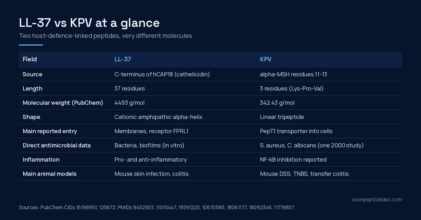
Short answer: LL-37 and KPV are both discussed as “host defence” peptides, but they are not two versions of the same idea. LL-37 is a 37-residue, positively charged helix cut from the human cathelicidin precursor hCAP18; it attacks bacterial membranes directly and also signals to immune cells[1][2][3]. KPV is the three-residue C-terminal fragment of alpha-melanocyte-stimulating hormone (alpha-MSH, residues 11-13); in cell and mouse work it enters cells through the peptide transporter PepT1 and dampens NF-kB inflammatory signalling[4]. One is mostly studied as an antimicrobial with complicated immune effects, the other mostly as an anti-inflammatory with one older antimicrobial report[5].
Two very different molecules
The size gap is the first thing to understand. PubChem lists LL-37 (CID 16198951, INN ropocamptide) at 4493 g/mol with the formula C205H340N60O53, and KPV (CID 125672, “MSH (11-13)”) at 342.43 g/mol with the formula C16H30N4O4. LL-37 is more than thirteen times heavier. That matters at the bench: LL-37 behaves like a small protein (it folds, self-associates and sticks to surfaces), while KPV behaves like a small, water-soluble metabolite.
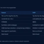
| Field | LL-37 | KPV |
|---|---|---|
| PubChem CID | 16198951 | 125672 |
| CAS (PubChem synonym) | 154947-66-7 | 67727-97-3 |
| Molecular formula | C205H340N60O53 | C16H30N4O4 |
| Molecular weight | 4493 g/mol | 342.43 g/mol |
| Sequence | LLGDFFRKSKEKIGKEFKRIVQRIKDFLRNLVPRTES | Lys-Pro-Val (KPV) |
| Parent molecule | hCAP18, the only human cathelicidin[1] | alpha-MSH (residues 11-13)[5] |
| Structure | Amphipathic alpha-helix, helicity depends on conditions[2] | Linear tripeptide |
| Best-described entry route | Membrane binding; FPRL1 receptor on leukocytes[6][3] | PepT1 di/tripeptide transporter[4] |
Identity values above are from PubChem and are not certificate-of-analysis values for any lot.
Where each one comes from
LL-37. The human cathelicidin was first predicted from a bone-marrow cDNA clone in 1995 as FALL-39, a cysteine-free peptide unlike the disulfide-bonded defensins; the synthetic peptide was active against E. coli and B. megaterium[7]. When the mature peptide was isolated from granulocytes, it turned out to be LL-37[1]. In neutrophils the holoprotein hCAP18 is cleaved to LL-37 after exocytosis, and proteinase 3 was the azurophil-granule protease responsible[8]. A separate 1995 cloning of human CAP18 showed that the C-terminal 37-residue domain binds lipopolysaccharide (LPS), inhibited LPS-induced nitric oxide release from macrophages and protected mice from LPS lethality[9].
KPV. KPV is not cut from a dedicated precursor in the way LL-37 is. It is simply the last three residues of alpha-MSH (SYSMEHFRWGKPV). A 2008 review explains why it attracted attention: alpha-MSH has broad anti-inflammatory effects in animal models, but it also drives pigmentation, and KPV keeps anti-inflammatory activity without the pigmentary action[10]. Our KPV vs BPC-157 comparison covers that melanocortin background in more depth.
Antimicrobial evidence: deep for LL-37, thin for KPV
LL-37’s antibacterial activity has been measured many ways. In a 1998 study its activity depended on its helical conformation: the minimal inhibitory concentration against E. coli was 5 µM, but at 13-25 µM it was also cytotoxic to several eukaryotic cell types, and human serum inhibited both the antibacterial and the cytotoxic activity[2]. Membrane work points to a detergent-like “carpet” action on negatively charged (bacterial-type) membranes, with possible pore formation in zwitterionic (mammalian-type) membranes, which helps explain why its cytotoxicity is not cell-selective[6].
Two further findings show the range. Against Pseudomonas aeruginosa, LL-37 blocked biofilm formation in vitro at 0.5 µg/mL, far below its growth-inhibiting concentration of 64 µg/mL, by reducing attachment and acting on quorum-sensing systems[11]. And in mice, genetic deletion of the cathelicidin gene (the mouse version is CRAMP) left animals less protected against necrotic skin infection by Group A Streptococcus[12], which is in vivo evidence that the cathelicidin system matters for host defence, not just test-tube killing.
For KPV the antimicrobial record rests mainly on one 2000 paper. There, alpha-MSH and KPV inhibited Staphylococcus aureus colony formation and reduced viability and germ-tube formation of Candida albicans over a broad concentration range; the effect appeared to involve increased cAMP in the yeast, and the peptides did not reduce killing by human neutrophils in vitro[5]. That is a useful starting point, but it is a far smaller evidence base than the LL-37 literature, and most later KPV work has focused on inflammation rather than microbial killing.
Inflammation: a double-edged peptide and a damper
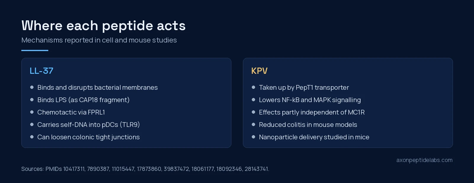
This is where the two separate most clearly. LL-37 is not simply anti-inflammatory. It is chemotactic for monocytes, neutrophils and T cells through the formyl peptide receptor-like 1 (FPRL1)[3], so it can recruit immune cells. In psoriasis research, LL-37 bound human self-DNA into condensed complexes that plasmacytoid dendritic cells took up, triggering Toll-like receptor 9 and type I interferon production[13], a mechanism proposed as a driver of autoimmune skin inflammation.
KPV studies point the other way. In human intestinal epithelial cells (Caco2-BBE, HT29-Cl.19A) and Jurkat T cells exposed to inflammatory cytokines, nanomolar KPV inhibited NF-kB and MAP kinase signalling and reduced inflammatory cytokine secretion, and those effects depended on PepT1-mediated uptake[4]. Because the parent hormone acts on melanocortin receptors, an obvious question is whether KPV does too. In mice carrying a non-functional melanocortin-1 receptor (MC1R), KPV still protected against DSS colitis, which the authors read as an effect at least partly independent of MC1R signalling[14].
The gut: where the comparison gets interesting
Both peptides have been studied in models of intestinal inflammation, and the results do not line up neatly.
- KPV in mouse colitis. KPV reduced DSS- and TNBS-induced colitis in mice and lowered inflammatory cytokine expression[4]. In a second group’s DSS and CD45RBhi transfer colitis models, KPV-treated mice recovered body weight sooner and showed fewer inflammatory infiltrates and lower myeloperoxidase activity[14]. Hyaluronic-acid-coated nanoparticles carrying KPV reached colonic epithelial cells and macrophages and reduced mucosal damage and TNF-alpha in a mouse ulcerative colitis model[15]. In a colitis-associated cancer model, KPV reduced tumour formation in wild-type mice but had no such effect in PepT1-knockout mice[16], a neat genetic test of the transporter route.
- LL-37 in the colon. The picture is mixed. In T84 human colonic epithelial cells, LL-37 caused a rapid, transient rise in permeability by driving endocytosis and lysosomal degradation of the tight-junction proteins occludin and claudin-2, at concentrations below those needed to kill microbes[17]. Another study found LL-37 forming complexes with bacterial DNA that were elevated in plasma and lesions of people with ulcerative colitis; the mouse equivalent (CRAMP-bacterial DNA) worsened mouse colitis, while interfering with the complex eased it[18].
A 2025 short review grouped cathelicidins and the alpha-MSH-derived KPV together among host defence peptides being explored for inflammatory bowel disease research, mainly because of reported effects on NF-kB and cytokine release[19]. The primary data above suggest those labels hide real differences: KPV results in mouse colitis have pointed in one direction, while LL-37 results depend heavily on context.
What this means for study design
- Serum and buffers change LL-37. Its helicity, antibacterial activity and cytotoxicity all shift with ionic conditions, and serum inhibited both activities in vitro[2]. Report media composition alongside any LL-37 result.
- Include a host-cell toxicity readout. LL-37 is cytotoxic to eukaryotic cells in the low micromolar range[2][6], so an “anti-inflammatory” signal in a cell assay should be checked against cell viability.
- Check PepT1 expression for KPV. KPV’s best-documented effects need PepT1[4][16]. A cell line with little PepT1 may show weak or no response for reasons that have nothing to do with the peptide’s quality.
- Confirm identity by mass. At about 342 versus about 4493 g/mol, the two are trivially separated by mass spectrometry, and LL-37 truncations (such as the N-terminally shortened FF-33 used in structure studies{c(“10417311”)}) are detectable the same way.
Research-grade material is listed as LL-37 and KPV 10mg; for the wider catalogue and lot documentation, you can buy lab-tested peptides in Canada. If you are comparing LL-37 with another immune peptide, see LL-37 vs thymosin alpha-1.
Frequently asked questions
Is KPV a fragment of LL-37?
No. KPV is residues 11-13 of alpha-melanocyte-stimulating hormone (Lys-Pro-Val). LL-37 is the 37-residue C-terminal peptide of the cathelicidin hCAP18. They come from unrelated precursors.
Which one has stronger antimicrobial evidence?
LL-37. Its antibacterial, anti-biofilm and membrane activity has been studied in many papers, and cathelicidin-deficient mice were less protected against Group A Streptococcus skin infection. KPV’s antimicrobial data come mainly from a single 2000 study against S. aureus and C. albicans.
Does KPV act through melanocortin receptors?
Not only. In cell studies its anti-inflammatory effects depended on uptake by the PepT1 transporter, and in mice lacking a functional MC1R it still reduced DSS colitis, suggesting a route at least partly independent of MC1R.
Is LL-37 anti-inflammatory?
It depends on context. LL-37 recruits immune cells through FPRL1, helps self-DNA activate plasmacytoid dendritic cells, and in colonic epithelial cells it increased permeability by degrading tight-junction proteins. Its immune effects are better described as modulatory than simply anti-inflammatory.
How can a lab tell the two apart?
By mass. PubChem lists LL-37 at 4493 g/mol and KPV at 342.43 g/mol, so mass spectrometry separates them easily. HPLC purity alone does not confirm identity.
References
- Gudmundsson GH, Agerberth B, Odeberg J et al. (1996). The human gene FALL39 and processing of the cathelin precursor to the antibacterial peptide LL-37 in granulocytes. Eur J Biochem. PMID: 8681941. doi: 10.1111/j.1432-1033.1996.0325z.x.
- Johansson J, Gudmundsson GH, Rottenberg ME et al. (1998). Conformation-dependent antibacterial activity of the naturally occurring human peptide LL-37. J Biol Chem. PMID: 9452503. doi: 10.1074/jbc.273.6.3718.
- De Yang, Chen Q, Schmidt AP et al. (2000). LL-37, the neutrophil granule- and epithelial cell-derived cathelicidin, utilizes formyl peptide receptor-like 1 (FPRL1) as a receptor to chemoattract human peripheral blood neutrophils, monocytes, and T cells. J Exp Med. PMID: 11015447. doi: 10.1084/jem.192.7.1069.
- Dalmasso G, Charrier-Hisamuddin L, Nguyen HT et al. (2008). PepT1-mediated tripeptide KPV uptake reduces intestinal inflammation. Gastroenterology. PMID: 18061177. doi: 10.1053/j.gastro.2007.10.026.
- Cutuli M, Cristiani S, Lipton JM et al. (2000). Antimicrobial effects of alpha-MSH peptides. J Leukoc Biol. PMID: 10670585. doi: 10.1002/jlb.67.2.233.
- Oren Z, Lerman JC, Gudmundsson GH et al. (1999). Structure and organization of the human antimicrobial peptide LL-37 in phospholipid membranes: relevance to the molecular basis for its non-cell-selective activity. Biochem J. PMID: 10417311.
- Agerberth B, Gunne H, Odeberg J et al. (1995). FALL-39, a putative human peptide antibiotic, is cysteine-free and expressed in bone marrow and testis. Proc Natl Acad Sci U S A. PMID: 7529412. doi: 10.1073/pnas.92.1.195.
- Sørensen OE, Follin P, Johnsen AH et al. (2001). Human cathelicidin, hCAP-18, is processed to the antimicrobial peptide LL-37 by extracellular cleavage with proteinase 3. Blood. PMID: 11389039. doi: 10.1182/blood.v97.12.3951.
- Larrick JW, Hirata M, Balint RF et al. (1995). Human CAP18: a novel antimicrobial lipopolysaccharide-binding protein. Infect Immun. PMID: 7890387. doi: 10.1128/iai.63.4.1291-1297.1995.
- Brzoska T, Luger TA, Maaser C et al. (2008). Alpha-melanocyte-stimulating hormone and related tripeptides: biochemistry, antiinflammatory and protective effects in vitro and in vivo, and future perspectives for the treatment of immune-mediated inflammatory diseases. Endocr Rev. PMID: 18612139. doi: 10.1210/er.2007-0027.
- Overhage J, Campisano A, Bains M et al. (2008). Human host defense peptide LL-37 prevents bacterial biofilm formation. Infect Immun. PMID: 18591225. doi: 10.1128/IAI.00318-08.
- Nizet V, Ohtake T, Lauth X et al. (2001). Innate antimicrobial peptide protects the skin from invasive bacterial infection. Nature. PMID: 11719807. doi: 10.1038/35106587.
- Lande R, Gregorio J, Facchinetti V et al. (2007). Plasmacytoid dendritic cells sense self-DNA coupled with antimicrobial peptide. Nature. PMID: 17873860. doi: 10.1038/nature06116.
- Kannengiesser K, Maaser C, Heidemann J et al. (2008). Melanocortin-derived tripeptide KPV has anti-inflammatory potential in murine models of inflammatory bowel disease. Inflamm Bowel Dis. PMID: 18092346. doi: 10.1002/ibd.20334.
- Xiao B, Xu Z, Viennois E et al. (2017). Orally Targeted Delivery of Tripeptide KPV via Hyaluronic Acid-Functionalized Nanoparticles Efficiently Alleviates Ulcerative Colitis. Mol Ther. PMID: 28143741. doi: 10.1016/j.ymthe.2016.11.020.
- Viennois E, Ingersoll SA, Ayyadurai S et al. (2016). Critical role of PepT1 in promoting colitis-associated cancer and therapeutic benefits of the anti-inflammatory PepT1-mediated tripeptide KPV in a murine model. Cell Mol Gastroenterol Hepatol. PMID: 27458604. doi: 10.1016/j.jcmgh.2016.01.006.
- Kilari G, Tran J, Blyth GAD et al. (2025). Human cathelicidin LL-37 rapidly disrupted colonic epithelial integrity. Biochim Biophys Acta Biomembr. PMID: 39837472. doi: 10.1016/j.bbamem.2025.184410.
- Duan Z, Fang Y, Sun Y et al. (2018). Antimicrobial peptide LL-37 forms complex with bacterial DNA to facilitate blood translocation of bacterial DNA and aggravate ulcerative colitis. Sci Bull (Beijing). PMID: 36658908. doi: 10.1016/j.scib.2018.09.014.
- Rodrigues JM, Ferreira Leal AP, Buccini DF et al. (2025). Host defense peptides as a new drug lead to a strategy for inflammatory bowel disease. Drug Discov Today. PMID: 41241376. doi: 10.1016/j.drudis.2025.104535.