
MOTS-c vs Humanin: Two Mitochondrial-Derived Peptides Compared
For laboratory research only. Not for human or animal use. This article gives no dosing, administration or therapeutic advice.
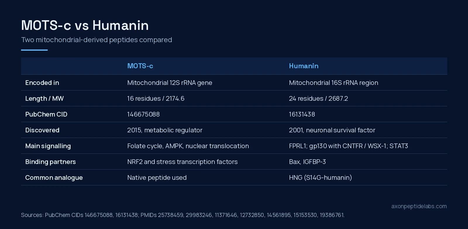
Short answer: MOTS-c and humanin are the two best-known mitochondrial-derived peptides, short peptides encoded by small open reading frames inside mitochondrial DNA. They come from neighbouring ribosomal RNA genes but have been studied for different reasons. Humanin, a 24-residue peptide from the 16S rRNA region, was discovered in 2001 as a factor that rescued neurons from death caused by familial Alzheimer’s disease genes and amyloid-beta[1][2]. MOTS-c, a 16-residue peptide from the 12S rRNA region, was reported in 2015 as a regulator of insulin sensitivity and metabolic homeostasis whose primary target appears to be skeletal muscle[3]. Put simply: humanin’s literature is built around cell survival and its receptors, MOTS-c’s around AMPK signalling, muscle and metabolic stress. The one direct cell-culture comparison we found, in human myotubes, gave them overlapping but not identical profiles[4].
Same genome, different neighbourhoods
Both peptides challenge the idea that the mitochondrial genome only encodes 13 proteins, 22 tRNAs and 2 rRNAs. The MOTS-c discovery paper says so directly: the identification of humanin as a signalling peptide encoded in mitochondrial DNA suggested further short open reading frames might exist, and MOTS-c was found inside the 12S rRNA gene[3]. Humanin sits in the 16S rRNA region, and an in silico search of that same region found six more candidates, named small humanin-like peptides (SHLPs), two of which shared humanin’s protective effects in vitro[2].
Humanin has a twist that MOTS-c lacks. The 2003 Bax study noted that the mitochondrial genome contains an open reading frame identical to the humanin sequence and that the mitochondrial version also binds and suppresses Bax, leading the authors to speculate that humanin arose in mitochondria and transferred to the nuclear genome[5]. When reading humanin papers, it is worth checking which version and which analogue was used.
Identity at a glance
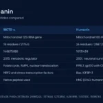
| Property | MOTS-c | Humanin |
|---|---|---|
| Full name | Mitochondrial open reading frame of the 12S rRNA-c | Humanin (HN) |
| Encoded in | Mitochondrial 12S rRNA gene | Mitochondrial 16S rRNA region (nuclear copies also exist) |
| Sequence | MRWQEMGYIFYPRKLR (16 residues) | MAPRGFSCLLLLTSEIDLPVKRRA (24 residues) |
| Molecular formula | C101H152N28O22S2 | C119H204N34O32S2 |
| Molecular weight | 2174.6 g/mol | 2687.2 g/mol |
| CAS (PubChem synonym) | 1627580-64-6 | 330936-69-1 |
| PubChem CID | 146675088 | 16131438 |
| Common research analogue | None widely used | HNG (S14G-humanin) |
Formula, weight and CAS values come from the linked PubChem records; the humanin sequence is read from PubChem’s systematic name. A practical point for lab work: much of the in vivo humanin literature uses the more potent glycine-14 analogue HNG rather than native humanin[6][4], whereas most MOTS-c studies use the native 16-residue peptide.
Humanin: a survival peptide with several receptors
Discovery in an Alzheimer’s screen
Humanin was found by functional expression screening for genes that protect neurons. The cDNA encoded a short secreted polypeptide that abolished neuronal death caused by several familial Alzheimer’s disease genes and by amyloid-beta, but not death caused by Q79 or SOD1 mutants, and the rescue depended on its primary structure[1].
Intracellular partners
Two intracellular partners were reported in 2003. Humanin bound Bax, blocked its association with isolated mitochondria and suppressed cytochrome c release in vitro[5]. Separately, humanin bound IGFBP-3 at the binding protein’s 18-residue heparin-binding domain; a single F6A substitution abolished binding, and the interaction could be either synergistic or antagonistic depending on the cell type[7].
Cell-surface receptors
Humanin also acts from outside the cell. It induced chemotaxis of mononuclear phagocytes through the G protein-coupled receptor FPRL1 (FPR2 in mice), a receptor amyloid-beta also uses, and blocked amyloid-beta’s toxicity in neuroblast cells[8]. A 2009 study added a second, cytokine-type route: gp130 was essential for humanin neuroprotection, and CNTF receptor alpha and the IL-27 receptor subunit WSX-1 were involved in humanin binding to neuronal cells[9]. Having both a GPCR route and a gp130 route is unusual for a 24-residue peptide and is one reason humanin papers report such varied effects.
Metabolism and ageing
In rodent clamp studies, humanin infused into the brain improved overall insulin sensitivity through hypothalamic STAT-3 signalling, and intravenous humanin derivatives reproduced the effect[10]. Circulating humanin declines with age in mice and humans; long-lived GH-deficient Ames mice had higher levels, short-lived GH-transgenic mice lower levels, and GH or IGF-I treatment reduced circulating humanin in mice and people[11]. In C. elegans, humanin overexpression extended lifespan in a daf-16/FOXO-dependent way, and children of centenarians had higher circulating humanin than age-matched controls[6].
MOTS-c: a mitochondrial signal to muscle and nucleus
AMPK and the folate cycle
The 2015 discovery paper reported that MOTS-c inhibits the folate cycle and its tethered de novo purine biosynthesis, leading to AMPK activation, with skeletal muscle as its apparent primary target organ. In mice, MOTS-c treatment prevented age-dependent and high-fat-diet-induced insulin resistance[3].
Moving into the nucleus
Under metabolic stress, MOTS-c translocated to the nucleus in an AMPK-dependent way, regulated a broad range of genes in response to glucose restriction, including antioxidant response element genes, and interacted with stress-responsive transcription factors such as NRF2[12]. This is the clearest mechanistic difference from humanin: MOTS-c is described as a mitochondria-to-nucleus messenger, humanin mainly as a ligand for membrane receptors and binding partner of apoptosis proteins.
Exercise and physical capacity in mice
MOTS-c enhanced physical performance in young, middle-aged and old mice, and in humans exercise induced endogenous MOTS-c expression in skeletal muscle and in circulation[13]. Human association data add nuance: plasma MOTS-c was similar in lean and obese adults in a 20-person study, though it correlated with insulin-sensitivity indices in lean participants[14].
A natural variant with less activity
An East Asian mitochondrial DNA variant, m.1382A>C, changes lysine 14 to glutamine (K14Q). In a meta-analysis of three cohorts totalling 27,527 people, men carrying the C allele had a higher prevalence of type 2 diabetes; in high-fat-fed male mice, native MOTS-c improved glucose tolerance but K14Q-MOTS-c did not[15]. A follow-up in Japanese cohorts found that C-allele carriers had a higher proportion of fast-twitch MHC-IIx fibres and that the allele was more frequent in sprint and power athletes than in endurance athletes[16].
Where the two peptides meet
Three studies put MOTS-c and humanin in the same frame:
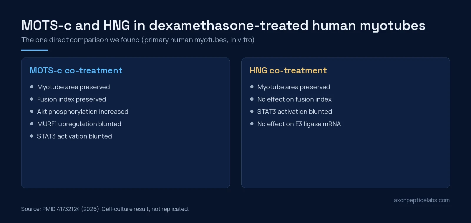
- Human myotubes. In primary human skeletal myotubes exposed to dexamethasone, MOTS-c co-treatment preserved myotube area and fusion index, increased Akt phosphorylation and blunted MURF1 upregulation and STAT3 activation. HNG preserved myotube area and blunted STAT3 activation but had no effect on fusion index or E3 ligase mRNA[4]. That is a cell-culture result, but it is the only direct comparison we found.
- Athletes. In serum from 30 non-athletes and 75 professional athletes, athletes had lower MOTS-c and higher humanin than sedentary controls, and the two levels were highly correlated in athletes but not in controls[17]. This is a pilot, cross-sectional study.
- The SHLP family. The humanin-region peptides SHLP2 and SHLP3 reduced apoptosis and reactive oxygen species and improved mitochondrial metabolism in vitro, circulating SHLP2, like humanin, decreased with age[2]. MOTS-c, from the 12S region, is not part of that family.
Side-by-side research comparison
| Question | MOTS-c | Humanin |
|---|---|---|
| Discovered | 2015, metabolic screen of mtDNA open reading frames[3] | 2001, neuronal survival screen[1] |
| Main signalling described | Folate cycle inhibition, AMPK activation; nuclear translocation[3][12] | FPRL1; gp130 with CNTFR and WSX-1; STAT3[8][9] |
| Intracellular partners | NRF2 and other stress-responsive transcription factors[12] | Bax, IGFBP-3[5][7] |
| Main tissue focus | Skeletal muscle[3][13] | Neurons, then metabolism and ageing[1][10] |
| Natural variant studied | K14Q (m.1382A>C)[15] | Analogue HNG used experimentally[6] |
| Change with age | Age-dependent physical decline studied in mice[13] | Circulating levels decline with age[11] |
Practical notes for study design
Three details from the primary papers matter when planning work with either peptide. First, decide whether the question is about the native peptide or an analogue: the humanin field moved early to HNG and other potent derivatives[10][6], so a protocol that uses native humanin is not directly comparable with much of the published work. Second, state the model. MOTS-c’s metabolic effects were first described in mice and in cultured cells[3], its nuclear translocation in cells under glucose restriction[12], and its human data are measurements of circulating or muscle levels[13][14]. Third, genotype can matter. The K14Q variant shows that a single residue change can blunt MOTS-c activity[15], and similar single-residue effects are documented for humanin, where the F6A substitution removed binding to IGFBP-3[7].
What the evidence does not show
- No human intervention data that settle anything. The human data for both peptides are associations: circulating levels in obesity, ageing, centenarian families or athletes[14][11][6][17]. None of this shows what giving either peptide does in people, and we found no FDA approval listing for either.
- Mouse and worm results do not transfer automatically. Lifespan extension was shown in C. elegans[6], and the physical-capacity and insulin findings come from mice and rats[13][3][10].
- Analogues are not the parent peptide. Many humanin results use HNG or other derivatives[10][6]. Results with an analogue should not be read as results with native humanin.
- Reviews are broader than the data. A 2023 systematic review presents humanin as a candidate for strategies against ageing processes such as cardiovascular disease, neurodegeneration and cancer[18]. That describes a research direction, not established effects.
- The direct comparison is a single in vitro study. The myotube paper[4] has not been replicated, and no animal study has compared the two head to head.
Sourcing MOTS-c for research
Axon carries MOTS-c 10 mg as a lyophilised vial and as a MOTS-c 10 mg pen; we do not stock humanin. Our COA library lists MOTS-C 10mg pen + vial, batch MP-MOTS-10-041626, 99.89% purity, analysed 28 APR 2026 by testides.com. One report covers both formats, so check the batch number on the label against the report. For background, see our MOTS-c research overview and the SS-31 vs MOTS-c comparison. Researchers who want to buy lab-tested peptides in Canada for mitochondrial-peptide work should note that MOTS-c is the better-characterised choice for AMPK and muscle models, while humanin questions sit closer to apoptosis and receptor biology.
Frequently asked questions
Where are MOTS-c and humanin encoded?
Both come from short open reading frames in mitochondrial DNA. MOTS-c is encoded in the 12S rRNA gene; humanin is in the 16S rRNA region, which also contains six small humanin-like peptides.
What is the main mechanistic difference?
MOTS-c is described as inhibiting the folate cycle, activating AMPK and moving into the nucleus under metabolic stress. Humanin acts through receptors such as FPRL1 and a gp130-containing complex and binds Bax and IGFBP-3.
What is HNG?
HNG is S14G-humanin, a more potent analogue with glycine at position 14. Many in vivo and cell studies use HNG rather than native humanin.
Have MOTS-c and humanin been compared directly?
One 2026 study in primary human myotubes exposed to dexamethasone compared MOTS-c with HNG. MOTS-c preserved myotube area and fusion index; HNG preserved area but not fusion index.
What is the MOTS-c K14Q variant?
It is an East Asian mitochondrial DNA variant, m.1382A>C, that swaps lysine 14 for glutamine. The K14Q peptide showed less insulin-sensitising activity in mice and cells.
References
- Hashimoto Y, Niikura T, Tajima H et al. (2001). A rescue factor abolishing neuronal cell death by a wide spectrum of familial Alzheimer’s disease genes and Abeta. Proc Natl Acad Sci U S A. PMID: 11371646. doi: 10.1073/pnas.101133498.
- Cobb LJ, Lee C, Xiao J et al. (2016). Naturally occurring mitochondrial-derived peptides are age-dependent regulators of apoptosis, insulin sensitivity, and inflammatory markers. Aging (Albany NY). PMID: 27070352. doi: 10.18632/aging.100943.
- Lee C, Zeng J, Drew BG et al. (2015). The mitochondrial-derived peptide MOTS-c promotes metabolic homeostasis and reduces obesity and insulin resistance. Cell Metab. PMID: 25738459. doi: 10.1016/j.cmet.2015.02.009.
- Elhusseiny R, Ihsan M, Bellefroid T et al. (2026). Mitochondrial-derived peptides MOTS-c and humanin attenuate dexamethasone-induced atrophy in human skeletal muscle cells. Physiol Rep. PMID: 41732124. doi: 10.14814/phy2.70791.
- Guo B, Zhai D, Cabezas E et al. (2003). Humanin peptide suppresses apoptosis by interfering with Bax activation. Nature. PMID: 12732850. doi: 10.1038/nature01627.
- Yen K, Mehta HH, Kim SJ et al. (2020). The mitochondrial derived peptide humanin is a regulator of lifespan and healthspan. Aging (Albany NY). PMID: 32575074. doi: 10.18632/aging.103534.
- Ikonen M, Liu B, Hashimoto Y et al. (2003). Interaction between the Alzheimer’s survival peptide humanin and insulin-like growth factor-binding protein 3 regulates cell survival and apoptosis. Proc Natl Acad Sci U S A. PMID: 14561895. doi: 10.1073/pnas.2135111100.
- Ying G, Iribarren P, Zhou Y et al. (2004). Humanin, a newly identified neuroprotective factor, uses the G protein-coupled formylpeptide receptor-like-1 as a functional receptor. J Immunol. PMID: 15153530. doi: 10.4049/jimmunol.172.11.7078.
- Hashimoto Y, Kurita M, Aiso S et al. (2009). Humanin inhibits neuronal cell death by interacting with a cytokine receptor complex or complexes involving CNTF receptor alpha/WSX-1/gp130. Mol Biol Cell. PMID: 19386761. doi: 10.1091/mbc.e09-02-0168.
- Muzumdar RH, Huffman DM, Atzmon G et al. (2009). Humanin: a novel central regulator of peripheral insulin action. PLoS One. PMID: 19623253. doi: 10.1371/journal.pone.0006334.
- Lee C, Wan J, Miyazaki B et al. (2014). IGF-I regulates the age-dependent signaling peptide humanin. Aging Cell. PMID: 25040290. doi: 10.1111/acel.12243.
- Kim KH, Son JM, Benayoun BA et al. (2018). The Mitochondrial-Encoded Peptide MOTS-c Translocates to the Nucleus to Regulate Nuclear Gene Expression in Response to Metabolic Stress. Cell Metab. PMID: 29983246. doi: 10.1016/j.cmet.2018.06.008.
- Reynolds JC, Lai RW, Woodhead JST et al. (2021). MOTS-c is an exercise-induced mitochondrial-encoded regulator of age-dependent physical decline and muscle homeostasis. Nat Commun. PMID: 33473109. doi: 10.1038/s41467-020-20790-0.
- Cataldo LR, Fernández-Verdejo R, Santos JL et al. (2018). Plasma MOTS-c levels are associated with insulin sensitivity in lean but not in obese individuals. J Investig Med. PMID: 29593067. doi: 10.1136/jim-2017-000681.
- Zempo H, Kim SJ, Fuku N et al. (2021). A pro-diabetogenic mtDNA polymorphism in the mitochondrial-derived peptide, MOTS-c. Aging (Albany NY). PMID: 33468709. doi: 10.18632/aging.202529.
- Kumagai H, Natsume T, Kim SJ et al. (2022). The MOTS-c K14Q polymorphism in the mtDNA is associated with muscle fiber composition and muscular performance. Biochim Biophys Acta Gen Subj. PMID: 34728329. doi: 10.1016/j.bbagen.2021.130048.
- Alser M, Ramanjaneya M, Rizwana Anwardeen N et al. (2022). The Effect of Chronic Endurance Exercise on Serum Levels of MOTS-c and Humanin in Professional Athletes. Rev Cardiovasc Med. PMID: 39077591. doi: 10.31083/j.rcm2305181.
- Coradduzza D, Congiargiu A, Chen Z et al. (2023). Humanin and Its Pathophysiological Roles in Aging: A Systematic Review. Biology (Basel). PMID: 37106758. doi: 10.3390/biology12040558.